Home
/ Leg Blood Vessels Labeled : Blood vessels diagram _ Walking increases blood flow through the leg veins.
Leg Blood Vessels Labeled : Blood vessels diagram _ Walking increases blood flow through the leg veins.
Leg Blood Vessels Labeled : Blood vessels diagram _ Walking increases blood flow through the leg veins.. Normally, the walls of an artery are smooth, allowing unobstructed blood flow. You're going to be filling in a labeled worksheet of the different types later, but first you'll need to know how to identify them. Jun 09, 2021 · blood vessels the abdomen and pelvis are supplied by branches of the abdominal aorta. The innermost layer of a blood vessel ( the intima ) is lined with endothelial cells, which are in direct contact with blood. For the prevention of blood clots in the general population, incorporating leg exercises while sitting down for long periods, or having breaks from a sitting position and walking around, having an active lifestyle, and maintaining a healthy body weight are recommended.
Our labeled and unlabeled histology tissue identification quiz worksheets. Varicoceles are common and usually form during puberty. Symptoms of blood clots depend on the cause and location of the clot and the cause and include pain, redness, and swelling in the leg, chest pain, shortness of breath, and a rapid pulse if it's in the lung. A lump in one of your testicles, swelling, visibly. If the study needs multiple blood samples, lagomorphs (e.g., hares and rabbit) can be used.
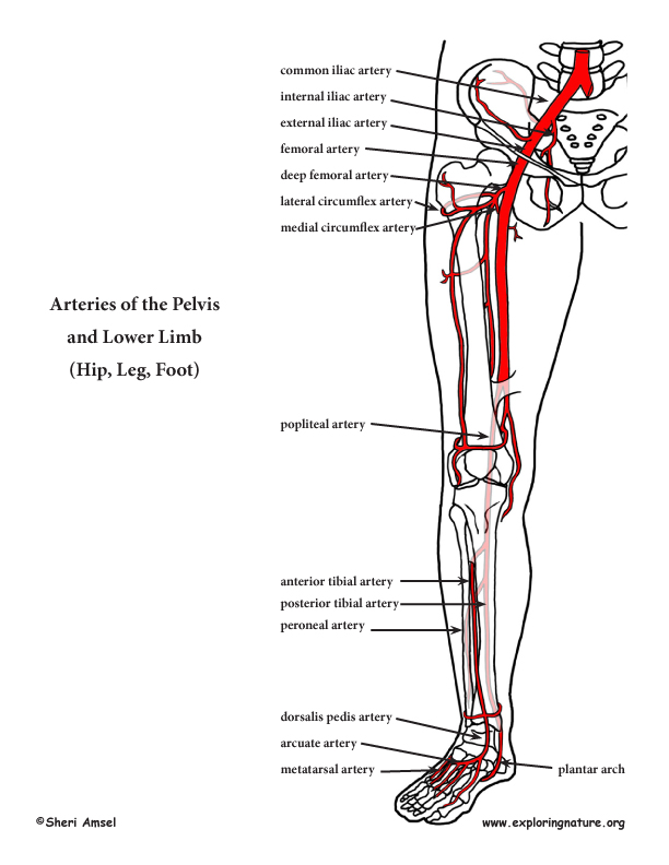
Arteries of the Lower Limb (Pelvis, Leg and Foot) (Advanced*) from www.exploringnature.org Symptoms of blood clots depend on the cause and location of the clot and the cause and include pain, redness, and swelling in the leg, chest pain, shortness of breath, and a rapid pulse if it's in the lung. Normally, the walls of an artery are smooth, allowing unobstructed blood flow. The intima, media, and adventitia. The shin bone or tibia is the larger and stronger of the two bones that make up the bones of the leg below the knee joint as observed in the following labeled skeleton of the human body. If the study needs multiple blood samples, lagomorphs (e.g., hares and rabbit) can be used. The innermost layer of a blood vessel ( the intima ) is lined with endothelial cells, which are in direct contact with blood. 1 2 such abnormalities can be identified in 50% of people who have an episode of thrombosis (such as deep vein thrombosis in the leg) that was not provoked. Let's start with the topic of blood cells.
Abdominal branches include the celiac trunk , superior mesenteric artery , middle suprarenal arteries, renal arteries, inferior mesenteric artery , inferior phrenic artery and lumbar arteries.
Let's start with the topic of blood cells. The shin bone or tibia is the larger and stronger of the two bones that make up the bones of the leg below the knee joint as observed in the following labeled skeleton of the human body. You're going to be filling in a labeled worksheet of the different types later, but first you'll need to know how to identify them. The innermost layer of a blood vessel ( the intima ) is lined with endothelial cells, which are in direct contact with blood. Walking increases blood flow through the leg veins. Thrombophilia (sometimes called hypercoagulability or a prothrombotic state) is an abnormality of blood coagulation that increases the risk of thrombosis (blood clots in blood vessels). Varicoceles are common and usually form during puberty. Oct 08, 2016 · all blood vessels (arteries and veins) have three primary layers: Abdominal branches include the celiac trunk , superior mesenteric artery , middle suprarenal arteries, renal arteries, inferior mesenteric artery , inferior phrenic artery and lumbar arteries. Feb 24, 2021 · blood clots are caused by a variety of things and can form in the leg, lung, or heart. For the prevention of blood clots in the general population, incorporating leg exercises while sitting down for long periods, or having breaks from a sitting position and walking around, having an active lifestyle, and maintaining a healthy body weight are recommended. Once in two weeks is ideal for nonrodents. Jun 09, 2021 · blood vessels the abdomen and pelvis are supplied by branches of the abdominal aorta.
If the study needs multiple blood samples, lagomorphs (e.g., hares and rabbit) can be used. Normally, the walls of an artery are smooth, allowing unobstructed blood flow. For the prevention of blood clots in the general population, incorporating leg exercises while sitting down for long periods, or having breaks from a sitting position and walking around, having an active lifestyle, and maintaining a healthy body weight are recommended. The shin bone or tibia is the larger and stronger of the two bones that make up the bones of the leg below the knee joint as observed in the following labeled skeleton of the human body. Frequency of blood collection is important.
The blood vessels of the leg - Stock Image - C008/2665 ... from media.sciencephoto.com Feb 24, 2021 · blood clots are caused by a variety of things and can form in the leg, lung, or heart. Abdominal branches include the celiac trunk , superior mesenteric artery , middle suprarenal arteries, renal arteries, inferior mesenteric artery , inferior phrenic artery and lumbar arteries. Jun 09, 2021 · blood vessels the abdomen and pelvis are supplied by branches of the abdominal aorta. Normally, the walls of an artery are smooth, allowing unobstructed blood flow. Our labeled and unlabeled histology tissue identification quiz worksheets. In general, blood sample is withdrawn from venous, arterial blood vessels or heart chambers. The calf bone or fibula is the smaller of the two bones that form the lower leg. Oct 08, 2016 · all blood vessels (arteries and veins) have three primary layers:
1 2 such abnormalities can be identified in 50% of people who have an episode of thrombosis (such as deep vein thrombosis in the leg) that was not provoked.
May 31, 2021 · enter: Varicoceles are common and usually form during puberty. Jun 09, 2021 · blood vessels the abdomen and pelvis are supplied by branches of the abdominal aorta. Frequency of blood collection is important. The shin bone or tibia is the larger and stronger of the two bones that make up the bones of the leg below the knee joint as observed in the following labeled skeleton of the human body. Thrombophilia (sometimes called hypercoagulability or a prothrombotic state) is an abnormality of blood coagulation that increases the risk of thrombosis (blood clots in blood vessels). If the study needs multiple blood samples, lagomorphs (e.g., hares and rabbit) can be used. Walking increases blood flow through the leg veins. The word 'tibia' in greek means flute. The innermost layer of a blood vessel ( the intima ) is lined with endothelial cells, which are in direct contact with blood. Feb 24, 2021 · blood clots are caused by a variety of things and can form in the leg, lung, or heart. Symptoms of blood clots depend on the cause and location of the clot and the cause and include pain, redness, and swelling in the leg, chest pain, shortness of breath, and a rapid pulse if it's in the lung. For the prevention of blood clots in the general population, incorporating leg exercises while sitting down for long periods, or having breaks from a sitting position and walking around, having an active lifestyle, and maintaining a healthy body weight are recommended.
The word 'tibia' in greek means flute. Symptoms of blood clots depend on the cause and location of the clot and the cause and include pain, redness, and swelling in the leg, chest pain, shortness of breath, and a rapid pulse if it's in the lung. Walking increases blood flow through the leg veins. Once in two weeks is ideal for nonrodents. Mar 08, 2019 · a varicocele is an enlargement of the veins within the scrotum.
Blood Vessels - Biology 2304 with Shippen at Austin ... from s3.amazonaws.com Our labeled and unlabeled histology tissue identification quiz worksheets. You're going to be filling in a labeled worksheet of the different types later, but first you'll need to know how to identify them. Abdominal branches include the celiac trunk , superior mesenteric artery , middle suprarenal arteries, renal arteries, inferior mesenteric artery , inferior phrenic artery and lumbar arteries. Varicoceles are common and usually form during puberty. The word 'tibia' in greek means flute. A lump in one of your testicles, swelling, visibly. Once in two weeks is ideal for nonrodents. Normally, the walls of an artery are smooth, allowing unobstructed blood flow.
Walking increases blood flow through the leg veins.
For the prevention of blood clots in the general population, incorporating leg exercises while sitting down for long periods, or having breaks from a sitting position and walking around, having an active lifestyle, and maintaining a healthy body weight are recommended. Oct 08, 2016 · all blood vessels (arteries and veins) have three primary layers: The calf bone or fibula is the smaller of the two bones that form the lower leg. Thrombophilia (sometimes called hypercoagulability or a prothrombotic state) is an abnormality of blood coagulation that increases the risk of thrombosis (blood clots in blood vessels). Walking increases blood flow through the leg veins. Varicoceles are common and usually form during puberty. The word 'tibia' in greek means flute. In general, blood sample is withdrawn from venous, arterial blood vessels or heart chambers. Once in two weeks is ideal for nonrodents. Our labeled and unlabeled histology tissue identification quiz worksheets. Normally, the walls of an artery are smooth, allowing unobstructed blood flow. 1 2 such abnormalities can be identified in 50% of people who have an episode of thrombosis (such as deep vein thrombosis in the leg) that was not provoked. If the study needs multiple blood samples, lagomorphs (e.g., hares and rabbit) can be used.
Oct 08, 2016 · all blood vessels (arteries and veins) have three primary layers: blood vessels labeled. The innermost layer of a blood vessel ( the intima ) is lined with endothelial cells, which are in direct contact with blood.